Images showing the complexity of the human body have been released as part of an exhibition showing cutting edge imaging techniques. The University of Edinburgh outdoor exhibition shows how these techniques are contributing to medical research.
Professor Joanna Wardlaw, Director of Edinburgh Imaging and the Brain Research Imaging Centre, said: "Advances in imaging technologies – such as MRI, MR-PET and microscopy – are vital for the continued discovery of new methods of diagnosing, monitoring and treating disease. This exhibition gives patients an insight to research that is happening right here in Edinburgh."
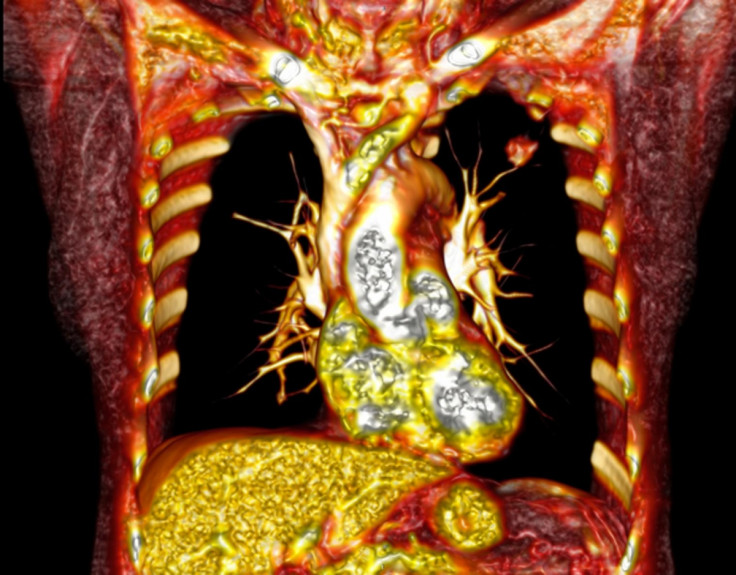
Images include a detailed look at the retina at the back of the eye, a 3D reconstruction of a patient's heart, aorta and coronary arteries and a magnified image of the small intestine.
The exhibition will take place at Little France and will run for five weeks from 3 September, 2015.
CT scans provide detailed 3D pictures of the insides of our bodies. This image reveals a red mass in the left lung that was found to be cancer. The patient later had surgery to remove this.Dr Saeed Mirsadraee at the Universe of Edinburgh
Cancer cells are continually growing and dividing, unlike normal cells which are strictly controlled. Scientists are studying what happens inside cells when they divide, identifying areas that can be targeted with drugs to stop them from dividing. Their aim is to develop treatments with fewer side effects than existing therapies.Dr Mar Carmena at the University of EdinburghMagnified image of the small intestine showing structures called Peyer's patches - the body's immune system.Dr David Donaldson and Prof Neil A Mabbott at the University of EdinburghThe wiring inside the brain of an ageing adult is captured by researchers probing why certain thinking skills slow as we get older. Coloured lines represent connections between areas of the brain's white matter. Yellow areas highlight connections that have been damaged as part of the ageing process.Dr Mark Bastin at the University of EdinburghMRIs provide the clearest pictures yet of unborn babies, allowing researchers to track their development. The brain, lung, kidney and bladder can be seen in the foetus on the left Dr Devasuda Anblagan at the University of EdinburghMagnified image showing human cells infected with influenza - the flu virus - shown in green.Matthew Turnbull at the University of EdinburghA firing brain cell (green) is blocked by dampening influences (red) and other surrounding cells (blue).Dr Nathalie Rochefort and Dr Janelle Pakan at the University of Edinburgh3D reconstruction of a patient's heart, aorta and coronary arteries created from CT scans.Dr Saeed Mirsadraee at the University of EdinburghImage shows a retina, with blood vessels highlighted in light blue. Measuring changes in retinal blood vessels could provide early warning of conditions like heart disease, diabetes and Alzheimer's Dr Tom MacGillivray at the University of Edinburgh