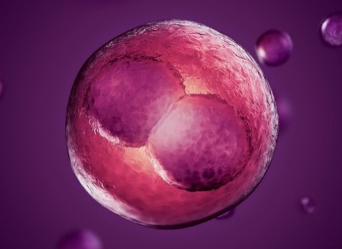
IVF Breakthrough as Time Lapse Imaging Triples Live Birth Rate
Fertility clinic develops method to work out the risk of a genetic abnormality in early embryos

A fertility clinic in Nottingham has developed a technique to increase the live birth rate, which has been described as the biggest breakthrough in IVF treatment for years.
Care Fertility has developed a time lapse imaging technique that allows doctors to establish which embryos are at the highest risk of having a genetic abnormality and therefore a decreased chance of success.
This means they are able to select the embryos that having the greatest chance of a live birth - Care researchers believe the live birth rate could triple (78%) using the technique.
Simon Fishel, managing director of the Care Fertility Group, said it was the "most exciting breakthrough in 30 years".
Stuart Lavery, director of IVF at Hammersmith Hospital, London, said: "Time lapse imaging of the early development of human embryos offers the exciting potential of a novel and non-invasive way of selecting the embryo with the greatest chance of implantation outside the womb."
In 2011, 48,147 women in the UK had IVF treatment. Of these, 17,041 had babies. Latest figures show that 25.6% of IVF treatments using a woman's own eggs result in a live birth.
Published in Reproductive BioMedicine Online, the study showed how abnormalities in five-day old human blastocysts can be identified by the rate at which they have developed, meaning the researchers could establish risk of genetic abnormality without a biopsy.
After an egg is fertilised, it may develop into an embryo over a period of about three days in a laboratory. A blastocyst is an embryo that has advanced to day five or six.
Around 40% of three day-old embryos will develop into blastocysts, meaning they are considered to be embryos with a higher chance of pregnancy.
Most embryos that fail to lead to a pregnancy do so because they have abnormal chromosomes. However, these abnormal embryos cannot be recognised without a biopsy.

The Care technique means embryologists can establish the risk of abnormality through imaging, rather than the using the invasive procedure.
Alison Campbell analysed blastocysts using time imaging and then took biopsies to test for abnormalities. She was then able to compare those with abnormal chromosome constitutions and with the blastocysts morphokinetic history (the biological process of it taking shape).
Embryos that were delayed in initiating blastocyst formation were more likely to have abnormalities, the authors found.
Campbell said: "This non-invasive model for the classification of chromosomal abnormality may be used to avoid selecting embryos with high risk of aneuploidy while selecting those with reduced risk."
Martin Johnson, editor of Reproductive BioMedicine Online, said: "Recently the world of IVF has become very excited by the use of time-lapse imaging of early human embryo development to follow the change of embryo morphology over time.
"The data can then be compared with the outcome after the embryos are transferred. The hope is that this morphokinetic analysis will enable reproductive specialists to predict more successfully those embryos most likely to generate pregnancies.
"The advantage of using morphokinetic analysis to predict outcome is its minimal invasiveness."
Markus Mongag, from the University Clinics of Heidelberg, added that the potential benefit of the Care approach was "obvious".
© Copyright IBTimes 2025. All rights reserved.


