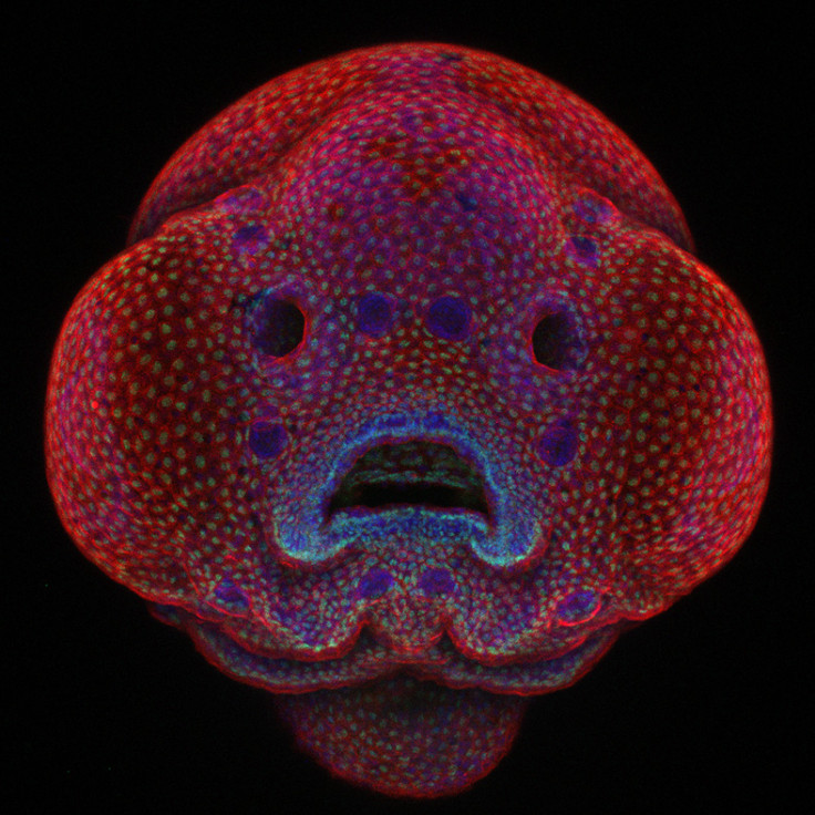
The winners of the annual Nikon Small World Photomicrography Competition have been revealed. Now in its 42nd year, the contest recognises excellence in photography taken under a microscope. First place was awarded to Oscar Ruiz PhD for his image of a four-day-old zebrafish embryo, bringing the world face-to-face with his research on facial development and cellular morphogenesis. Ruiz uses the zebrafish to study genetic mutations that lead to facial abnormalities such as cleft lip and palate in humans.
1st Place: Dr Oscar Ruiz, the University of Texas MD Anderson Cancer Center, Houston, Texas, US: Four-day-old zebrafish embryo – Confocal, 10xDr Oscar Ruiz
The judges were intrigued by Ruiz's innovative techniques to capture time-lapse images of the developing zebrafish face. Using the time-lapse as a guide, Ruiz is creating an atlas of the development of the zebrafish face. His group is tracking physical landmarks throughout development to create a series of metrics that can be used to accurately describe the cellular movements that occur during the normal development of the face. These metrics can then be used to identify abnormalities in the development of zebrafish harbouring specific genetic mutations identified in human patients. He hopes that these findings will help provide insight into the cellular and molecular mechanisms that are altered in patients with facial deformities.
The competition received more than 2,000 entries by scientists, photographers and hobbyists from 70 countries. Judges selected winners that exemplified artistic quality as well as exceptional scientific technique. In this gallery, IBTimes UK reveals the judges' top 20 . See more at the Nikon Small World site.
2nd Place: Douglas L Moore, University of Wisconsin – Stevens Point Museum of Natural History, Stevens Point, Wisconsin, USA: Polished slab of Teepee Canyon agate – Stereomicroscopy, 90xDouglas L Moore3rd Place: Rebecca Nutbrown University of Oxford, Nuffield Department of Clinical Neurosciences Oxford, United Kingdom: Culture of neurons (stained green) derived from human skin cells, and Schwann cells, a second type of brain cell (stained red) – Confocal/Immunofluorescence/iPSCs, 20xRebecca Nutbrown4th Place: Jochen Schroeder, Chiang Mai, Thailand: Butterfly proboscis – Image Stacking, 6.3xJochen Schroeder5th Place: Dr Igor Siwanowicz, Howard Hughes Medical Institute (HHMI), Janelia Research Campus, Ashburn, Virginia, USA: Front foot (tarsus) of a male diving beetle – Confocal, 100xDr Igor Siwanowicz6th Place: Marek Mis, Podlaskie, Poland: Air bubbles formed from melted ascorbic acid crystals – Polarized Light, 50xMarek Mis7th Place: Dr David Maitland, Feltwell, United Kingdom: Leaves of Selaginella (lesser club moss) – Differential Interference Contrast, 40xDr David Maitland8th Place: Samuel Silberman, Monoson Yahud, Israel: Wildflower stamens – Fiber Optic Illumination, 40xSamuel Silberman9th Place: Vin Kitayama and Sanae Kitayama, Vinsanchi Art Museum, Azumino, Nagano, Japan Espresso coffee crystals – Polarized LightVin Kitayama and Sanae Kitayams10th Place: Rogelio Moreno Gill, Panama, Panama: Frontonia (single-celled organism) showing ingested food, cilia, mouth and trichocysts – Differential Interference Contrast, 200xRogelio Moreno Gill11th Place: Francis Sneyers Brecht, Belgium: Scales of a butterfly wing underside (Vanessa atalanta) – Macroscopy, 10xFrancis Sneyers Brecht12th Place: Dr Dylan Burnette, Vanderbilt University School of Medicine Nashville, Tennessee, USA – Human HeLa cell undergoing cell division (cytokinesis). DNA (yellow), myosin II (blue) and actin filaments (red) – Structured Illumination, 9xDr Dylan Burnette13th Place: Walter Piorkowski, South Beloit, Illinois, USA: Poison fangs of a centipede (Lithobius erythrocephalus) – Fibre Optic Illumination/Image Stacking, 16xWalter Piorkowski14th Place: Dr Keunyoung Kim, University of California, San Diego, National Center for Microscopy and Imaging Research, La Jolla, California, USA: Mouse retinal ganglion cells – Fluorescence/Confocal 40xDr Keunyoung Kim15th Place: Geir Drange, Asker, Norway: Head section of an orange ladybird (Halyzia sedecimguttata) – Reflected Light/Focus Stacking, 10xGeir Drange16th Place: Stefano Barone, Diatom Shop, Palazzo Pignano, Italy: 65 fossil Radiolarians (zooplankton) carefully arranged by hand in Victorian style – Darkfield, 100xStefano Barone17th Place: Jose Almodovar, University of Puerto Rico, Mayaguez Campus, Biology Department, Mayaguez, Puerto Rico: Slime mould (Mixomicete) – Image Stacking/Reflected Light, 5xJose Almodovar18th Place: Pia Scanlon, Department of Agriculture and Food, Western Australia: Parts of wing-cover (elytron), abdominal segments and hind leg of a broad-shouldered leaf beetle (Oreina cacaliae) – Stereomicroscopy, Image Stacking, 40xPia Scanlon19th Place: Dr Gist F Croft, Lauren Pietilla, Stephanie Tse, Dr. Szilvia Galgoczi, Maria Fenner, Dr Ali H. Brivanlou, Rockefeller University, Brivanlou Laboratory New York, New York, USA: Human neural rosette primordial brain cells, differentiated from embryonic stem cells – Confocal, 10xDr Gist F Croft, Lauren Pietilla, Stephanie Tse, Dr. Szilvia Galgoczi, Maria Fenner, Dr Ali H. Brivanlou20th Place: Michael Crutchley, Haverfordwest, Pembrokeshire, United Kingdom: Fungi on cow dung – Darkfield, 30xMichael Crutchley
The Nikon Small World Popular Vote continues until 25 October. Go to the Nikon Small World site and take a look at all 95 finalists, then vote for your favourite.