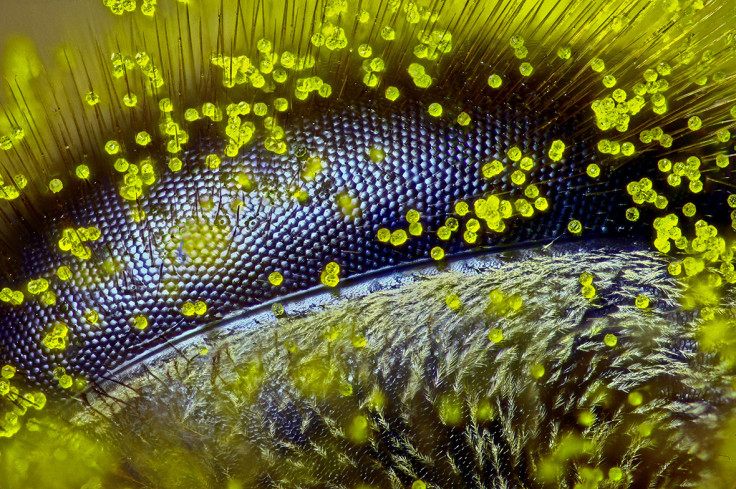
An incredible close-up image of dandelion pollen grains on a honey bee's eye has won the Nikon Small World 2015 photomicography competition. Ralph Grimm, an Australian high-school teacher and self-taught photomicrographer, says that as a former beekeeper, the subject matter is near and dear to his heart.
Colonies and bee populations continue to dwindle, and he hopes his image can serve as a voice for this rapidly disappearing insect that plays such a critical function in pollinating the world's crops. "In a way I feel as though this gives us a glimpse of the world through the eye of a bee," the Nikon competition winner said. "It's a subject of great sculptural beauty, but also a warning – that we should stay connected to our planet, listen to the little creatures like bees, and find a way to protect the earth that we all call home."
Judges were particularly impressed by the technique Grimm employed to capture this image stack, which included over four hours of careful work to mount the eye, set the focus increments, properly illuminate the eye and avoid peripheral smudging during the stacking process. The resulting image is a testament to Grimm's painstaking efforts.
1st Place: Ralph Claus Grimm from Jimboomba, Queensland, Australia. Eye of a honey bee (Apis mellifera) covered in dandelion pollen (120x). Reflected LightRalph Claus Grimm/Nikon Small World 2015
Grimm now joins the ranks of 37 other photomicrographers, artists and scientists from all over the world who have taken the top prize. "Each year we are blown away by the incredible quality and quantity of microscopic images submitted from all over the world, from scientists, artists, and photomicrographers of all levels and backgrounds. This year was certainly no exception," said Eric Flem, communications manager, Nikon Instruments. "Judges had their work cut out for them in narrowing down from such a rich pool of applicants, and we are so pleased with the results."
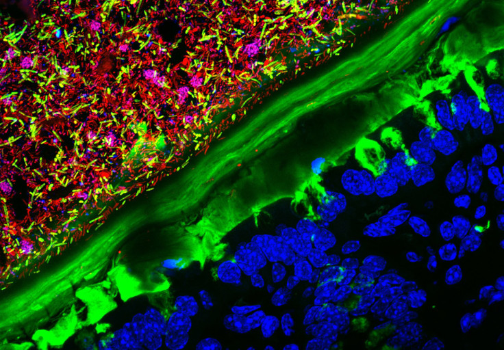
Kristen Earle, who has just completed a PhD in microbiology and immunology at Stanford, was placed second with her image of a mouse colon that has been colonised by human gut microbiota.
2nd Place: Kristen Earle, Gabriel Billings, KC Huang & Justin Sonnenburg from Stanford University School of Medicine, Department of Microbiology and Immunology Stanford, California, USA. Mouse colon colonised with human microbiota (63x). ConfocalKristen Earle, Gabriel Billings, KC Huang & Justin Sonnenburg/Nikon Small World 2015
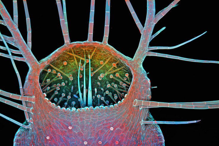
Third place went to Dr Igor Siwanowicz and his image of an entrance to the trap (or bladder) of Humped Bladderwort (Urticulatia gibba), a carnivorous freshwater plant. The bladderwort's trap is one of the most – if not the most – sophisticated plant organs in existence. Several elements of the bladder's construction are visible in the image, giving some insight into working of this tiny – only 1.5mm long – but elaborate suction trap.
3rd Place: Dr Igor Siwanowicz from Hughes Medical Institute (HHMI), Janelia Farm Research Campus, Leonardo Lab. Intake of a humped bladderwort (Utricularia gibba), a freshwater carnivorous plant (100x). ConfocalDr Igor Siwanowicz/Nikon Small World 2015
Now in its 41st year, Nikon Small World is widely regarded as the leading forum to recognise proficiency and excellence of photography taken under the microscope. To select the winners, competition judges analysed entries from all over the world covering subjects ranging from chemical compounds to up-close-and-personal looks at biological specimens. With more than 2,000 submissions spanning 83 countries, the 2015 Small World competition was tough. Entries had to display not only artistic quality but exceptional scientific technique. IBTimesUK presents the rest of the judges' top 20 ranked images.
4th Place: Daniel H Miller & Ethan S Sokol from Whitehead Institute for Biomedical Research, Massachusetts Institute of Technology, Department of Biology, Cambridge, Massachusetts, USA. Lab-grown human mammary gland organoid (100x). ConfocalDaniel H Miller & Ethan S Sokol/Nikon Small World 20155th Place: Dr Giorgio Seano & Dr Rakesh K Jain from Harvard Medical School, Massachusetts General Hospital, Edwin L Steele Laboratory for Tumour Biology, Boston, Massachusetts, USA. Live imaging of perfused vasculature in a mouse brain with glioblastoma. Optical Frequency Domain Imaging SystemDr Giorgio Seano & Dr Rakesh K Jain/Nikon Small World 20156th Place: Henri Koskinen from Helsinki, Finland. Spore capsule of a moss (Bryum sp). Reflected LightHenri Koskinen/Nikon Small World 20157th Place: Evan Darling from Memorial Sloan Kettering Cancer Centre, New York, New York, USA. Starfish imaged using confocal microscopy (10x). ConfocalEvan Darling/Nikon Small World 20158th Place: Dr Tomoko Yamazaki, National Institutes of Health (NIH), Bethesda, Maryland, USA. Nerves and blood vessels in a mouse ear skin (10x). ConfocalDr Tomoko Yamazaki/Nikon Small World 20159th Place: Dr Nathanael Prunet, California Institute of Technology and Dartmouth College, Department of Biology Pasadena, California, USA. Young buds of Arabidopsis (a flowering plant) (40x). ConfocalDr Nathanael Prunet/Nikon Small World 201510th Place: Ian Gardiner, Calgary, Alberta, Canada. Clam shrimp (Cyzicus mexicanus), live specimen (25x). Darkfield, Focus StackingIan Gardiner/Nikon Small World 201511th Place: Rogelio Moreno Gill, Panama. Fern sorus at varying levels of maturity (20x). Fluorescence, Image StackingRogelio Moreno Gill/Nikon Small World 201512th Place: Hannah Sheppard-Brennand, Southern Cross University, National Marine Science Centre Sydney, New South Wales, Australia. Developing sea mullet (Mugil cephalus) embryos (40x). BrightfieldHannah Sheppard-Brennand/Nikon Small World 201513th Place: Jose Almodovar, University of Puerto Rico (UPR), Mayaguez Campus, Biology Department Mayaguez, Puerto Rico, USA. Tentacles of a carnivorous plant (Drosera sp.) (20x). Image StackingJose Almodovar/Nikon Small World 201514th Place: Viktor Sykora, Charles University, First Faculty of Medicine Prague, Czech Republic. Australian grass (Austrostipa nodosa) seed (5x). DarkfieldViktor Sykora/Nikon Small World 201515th Place: Dr Heiti Paves, Tallinn University of Technology, Department of Gene Technology Tallinn, Estonia. Anther of a flowering plant (Arabidopsis thaliana) (20x). ConfocalDr Heiti Paves/Nikon Small World 201516th Place: Charles B Krebs, Charles Krebs Photography. Issaquah, Washington, USA. Feeding rotifers (Floscularia ringens) (50x) Darkfield.Charles B Krebs/Nikon Small World 201517th Place: Dr David Maitland, Feltwell, United Kingdom. Black witch-hazel (Trichodactylus crinitus) leaf producing crystals to defend against herbivores (100x). Differential Interference ContrastDr David Maitland/Nikon Small World 201518th Place: Roland Gross, Gruenen, Bern, Switzerland. Hairyback worm (Chaetonotus sp.) and algae (Micrasterias sp.) (400x). Differential Interference ContrastRoland Gross/Nikon Small World 201519th Place: Dr Richard R Kirby, Marine Biological Association, Plymouth, United Kingdom. Planktonic larva of a horseshoe worm (phoronid) (450x). DarkfieldDr Richard R Kirby/Nikon Small World 201520th Place: Frank Reiser, Nassau Community College, Department of Biology Garden City, New York, USA. Suction cups on the diving beetle (Dytiscus sp.) foreleg (50x). Image Stacking, PhotomergeFrank Reiser/Nikon Small World 2015
Top images from the 2015 Nikon Small World Competition will be exhibited in a full-colour calendar and through a US national museum tour. For additional information, please visit Nikon's website or follow @NikonSmallWorld on Twitter or Facebook.