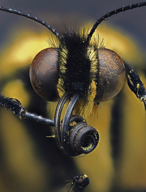
Twenty of the year's best scientific images have been chosen as finalists in the 2016 Wellcome Image Awards. This year's shortlist includes a map of the human brain's pathways, delicate scales on a moth's wing and a close-up of a butterfly's proboscis. The finalists were chosen by nine judges from all those acquired by the Wellcome Images picture library in the past year. The overall winner will be revealed at a ceremony on 15 March 2016.
Photomacrograph of the head of a swallowtail butterfly. Butterflies have two round compound eyes, which they use to see quick movements. From between the eyes extend two antennae, which are used as sensors, for example detecting smell and sniffing out a mate. They also have a long feeding tube (proboscis), which is curled up like a spring here, but it unrolls so the butterfly can use it like a straw to drink nectar from flowers. Swallowtail butterflies are widely distributed around the world and are often found in wetlands such as marshes and fens. They get their name from their characteristic hindwing extensions, which are reminiscent of a swallow’s tail. This image is 5 mm wide.
Daniel Saftner, Macroscopic Solutions
Pathways of nerve fibres in the brain of a young healthy adult (viewed from behind). Different parts of the brain communicate with each other through these nerve fibres, which are colour-coded here. Fibres connecting the left and right hemispheres are red, fibres travelling up and down connecting the brain and spinal cord are blue, and fibres running front to back are green. This image was created from virtual slices of the brain, from top to bottom, made using a type of magnetic resonance imaging (MRI) called diffusion imaging that tracks the direction and movement of water molecules. This information was then used to digitally reconstruct this network of connections. The width of this brain is 16.5 cm.
Alfred Anwander, Max Planck Institute for Human Cognitive and Brain Sciences
Thermal image of a healthy hand (left) and the hand of a person with Raynaud’s disease (right). Raynaud’s disease usually affects the hands and feet, causing less blood to flow to them when the person is cold, anxious or stressed. This makes the affected area turn pale and can also lead to numbness, pain, and pins and needles. These hands are from two different people, one male and one female. Both hands were put in cold water for 2 minutes before being imaged. The healthy hand then warmed considerably faster, and hotter areas are shown in yellow and orange. Colder areas are blue, black and purple. Thermal imaging detects infrared radiation or heat.
Matthew Clavey, Thermal Vision Research
Photograph of blisters on the arm of a girl with a black henna tattoo. Henna is commonly used to stain skin or hair orange-brown, but chemical dyes can be added to turn the colour black. These extra chemicals can cause allergic reactions and chemical burns. These blisters may lead to scarring and sometimes affect the natural colouring of the skin. The chemical responsible for these reactions is paraphenylenediamine, which is also widely used in permanent hair dyes – but its use in these products is strictly controlled. Temporary tattoos, which are drawn or painted onto the skin and then fade over time, are becoming increasingly popular.
Nicola Kelley, Cardiff and Vale University Hospital NHS Trust
Watercolour and ink illustration looking inside an Ebola virus particle. The virus is surrounded by a membrane (pink/purple) stolen from an infected cell. This is studded with proteins from the virus (turquoise) which extend outwards and look like trees rooted in the membrane. These proteins attach to the cells that the virus infects. A layer of proteins (blue) supports the membrane on the inside. Genetic information (RNA; yellow) is stored in a cylinder (nucleocapsid; green) in the centre of the virus. The Ebola virus, which first appeared in Africa in the mid-1970s, can cause serious illness and is often fatal. It can spread between people through direct contact with infected blood and other body fluids. Good hygiene, such as handwashing, is one important way to minimise the virus’s spread. This virus is approximately 100 nanometres (0.0001 mm) wide, which is 200 times smaller than many of the cells that it infects.
David S Goodsell, RCSB Protein Data Bank
A blockage (green) is highlighted in one of the blood vessels (red) in the neck of this person, just below the jaw. This blood vessel supplies blood to the brain. If it starts to clog, the inside of the artery can ‘fur up’. This furry patch can become unstable and burst, forming blood clots that can trigger a stroke. To allow treatment to reduce the risk of this, researchers are developing new ways of identifying these furry areas before they burst. This is being done with a combination of two simultaneous medical scans. Computed tomography (CT) uses X-rays to take virtual slices of the body to show the location of blood vessels and bones. Positron emission tomography (PET) uses radioactive markers to highlight any unstable furry patches inside arteries.
Nicholas Evans, University of Cambridge
3D image looking inside the back of a human eye. These tunnel-like structures are blood vessels which carry blood into the eye to provide it with the nutrition it needs. This layer of tissue (the choroid) is found at the back of the eyeball between the white of the eye and the retina, and is the major blood supply for the retina. The shape and structure of this tissue is unique in every person, like a fingerprint. This image was created using information from a 3D optical coherence tomography scan, using a new method for extracting and analysing the data. This type of imaging is being used to detect diseases such as glaucoma, diabetes, multiple sclerosis and age-related macular degeneration. Blood cells are not visible here as they are moving too quickly to be included. These tunnels or channels are approximately 100 micrometres (0.1 mm) tall.
Peter Maloca, University of Basel
Photograph of a newborn baby receiving light therapy in the Starlight Neonatal Unit at Barnet Hospital in London. This baby was born prematurely and has jaundice, a common condition where waste from the breakdown of red blood cells builds up in the blood, causing the skin and eyes to turn yellow. This waste, bilirubin, is normally removed by the liver, but in newborn babies the liver is not yet fully developed so cannot always do this efficiently. In a small number of cases jaundice may be a sign of an underlying health problem. This baby is being treated in a special incubator and lies under an ultraviolet light, eyes covered.
David Bishop, Royal Free Hospital, London
Fluorescein angiogram to show blood vessels inside a person’s eye. These blood vessels supply nutrients to the retina, the thin layer of light-sensitive tissue which lines the back of the inside of the eye and works a bit like film in a camera. Light enters the eye and is focused onto the retina, which then converts these images into electrical signals and sends them on to the brain to make sense of. If one of these blood vessels gets blocked or starts to leak, this can cause problems in the eye and may affect eyesight. To create an image like this fluorescent dye is first injected into a person’s arm. The dye then travels through the person’s veins and eventually through the blood vessels in the eye, where it highlights them so they can be photographed with a special type of camera. This image is roughly 20 mm wide.
Kim Baxter, Cambridge University Hospitals NHS Foundation Trust
Photograph of the UK’s only high-level isolation unit, at the Royal Free Hospital in London. A specially designed see-through tent is set up around the bed of a patient requiring treatment for a dangerous infectious disease such as viral haemorrhagic fever or Ebola. All air leaving the unit is cleaned, so the patient can be safely treated without putting other patients or staff at risk. This photograph was taken the day before William Pooley was admitted to the Royal Free in August 2014, after contracting Ebola in Sierra Leone. William worked as a nurse when the disease outbreak was at a critical stage and returned to Sierra Leone after making a full recovery. The high-level isolation unit is always on standby, ready to admit a patient at short notice.
David Bishop, Royal Free Hospital, London
Confocal micrograph of a small piece of human liver tissue put into a mouse with a damaged liver. Human liver cells (red/orange) and human blood vessels (green) in the new liver have grouped together and started to grow using blood (white) from the mouse to help. Development of blood vessels in organs like the liver has previously been very difficult, which has been a major barrier to scaling up small implants like these for medical use. The liver can regenerate itself but certain types of damage are irreversible, and there is a growing shortage of replacement organs. Researchers hope that one day implants like this could be used to repair livers damaged by liver disease, cirrhosis or cancer. The image is 1.1 mm wide.
Chelsea Fortin, Kelly Stevens and Sangeeta Bhatia, Koch Institute, © MIT
Photomacrograph of scales on a Madagascan sunset moth (Chrysiridia rhipheus). This is a large, colourful moth which flies during the day, while most other moths are only active at night. It is native to Madagascar and is often mistaken for a butterfly. As the wings move, they shimmer in the light and change colour, but these colours are an illusion. They come from light bouncing off the curved scales at different angles. The wings themselves hardly contain any colour pigment or dye. This image is 750 micrometres (0.75 mm) wide.
Mark R Smith, Macroscopic Solutions
Transmission electron micrograph of two rod-shaped bacteria sitting on an extremely thin sheet of graphene. This material is a sheet of carbon one atom thick, and has been described as a wonder material as it is one of the thinnest, strongest materials so far discovered and conducts electricity more efficiently than copper. The bacteria seen here were wrapped up in the graphene sheet by chance when non-sterile water was used in an experiment. Researchers are trying to stick different medicines to this material so they can be carried to the right place in the body when needed – for example, using antimicrobial drugs to kill bacteria or anticancer drugs to kill cancer cells. Each bacterium is approximately 2 micrometres (0.002 mm) long.
Izzat Suffian, Kuo-Ching Mei, Houmam Kafa and Khuloud T Al-Jamal, King’s College London
As babies and children develop, the structure of their bones changes. Each sphere shows bone from the backbone (the first lumbar vertebra, L1, in the lower back) of an infant at a different age. From left to right: 3 months before birth, just before birth, within the first year of life, 1.2 years old and 2.5 years old. These historical bones, from skeletal remains of children who died in the 19th century, were donated by the Royal College of Surgeons of England. Virtual X-ray slices of bone were taken and used to create a digital 3D model. Spheres of different sizes were virtually cut out from the model, to ensure that the same part of the bone was analysed in each case. The spheres are not to scale here, so that structural changes in the bone can be easily seen. From left to right they measure 2 mm, 3.5 mm, 5 mm, 10 mm and 8 mm wide.
Frank Acquaah
Clathrin is a protein found in cells which forms basket- or cage-like structures around small membrane sacs. These clathrin cages help bring molecules into the cell and then carry them around inside from one place to another. They also help sort through the molecules (which can include receptors and nutrients) so that different cargo is efficiently delivered to different destinations. Some disease-causing germs and toxins hijack this process and use it to infect cells. When the cage is not being used it breaks up into smaller pieces, which get recycled. Each building block of the cage is a triskelion, a pattern of three bent legs (dark blue) joined together with three short rods (light blue) attached. This digital illustration was created using scientific data (Protein Data Bank entry 1xi4) about the sequence, shape, size and fit of these building blocks and how they assemble into cages. Cages can form in different sizes, usually less than 200 nanometres (0.0002 mm) across. This particular cage is approximately 50 nanometres (0.00005 mm) across.
Maria Voigt, RCSB Protein Data Bank
Time-lapse confocal micrographs of a stem cell dividing in the brain of a zebrafish before it hatches. Starting at about the 8 o’clock position, this stem cell divides as you move clockwise through the stages to make two different cells: a nerve cell (on the outside, turning from purple to white) and another stem cell (on the inside, staying purple), which can itself go on and continue dividing. The sequence takes 9 hours for the two cells to separate and move apart. This type of unequal cell division ensures that both types of cell (nerve and stem cell) are made, as both are required for brain growth and development. The image is approximately 250 micrometres (0.25 mm) wide.
Paula Alexandre, University College London
Super-resolution micrograph of three parasites that can cause an infection called toxoplasmosis. You can catch these parasites (Toxoplasma gondii) by eating raw or undercooked meat or from contact with infected cat poo. They are dangerous to people with weak immune systems and to pregnant women, as a mother can pass the infection onto her unborn child. Here, DNA inside each parasite (blue/green) is surrounded by membrane (red) and protein (black). This image was created using a type of super-resolution microscopy called structured illumination microscopy. Each parasite measures 10 micrometres (0.01 mm).
Leandro Lemgruber, University of Glasgow
Cryogenic scanning electron micrograph of a single human stem cell. This type of stem cell has a natural ability to repair damaged tissue and can divide to produce some of the different cells found in the body. This cell is sitting in a mixture of chemicals designed to mimic its natural environment inside the body, so that researchers can better understand how it interacts with its surroundings. The stem cell is from inside the hip bone of a healthy person who donated some bone marrow to help treat patients who develop complications after receiving a marrow transplant. To create this image, the sample was first preserved by rapid freezing at cryogenic temperatures (lower than −150°C or −238°F). Electrons were then bounced off the surface of the frozen sample to provide information on its structure. The diameter of the cell is approximately 15 micrometres (0.015 mm).
Sílvia A Ferreira, Cristina Lopo and Eileen Gentleman, King’s College London
Confocal micrograph looking inside a cluster of leaves from a young maize (corn) plant. Each curled leaf is made up of lots of small cells (small green square and rectangle shapes), and inside each cell is a nucleus (orange circle), the part of the cell which stores genetic information. Maize is one of the most widely grown cereal crops in the world. It is used as a staple food, in livestock feed, and as a raw material – such as for processing into high-fructose corn syrup. Genetically modified maize crops are being grown to be resistant to pests and herbicides. This image is approximately 250 micrometres (0.25 mm) wide. Created in collaboration with Jim Haseloff and OpenPlant Cambridge.
Fernán Federici, Pontificia Universidad Católica de Chile and University of Cambridge
A preserved heart from an adult cow. It is made of four chambers through which blood flows: two upper chambers (where blood enters the heart) and two lower chambers (where blood exits the heart). Here, windows have been cut into three of these chambers to show what’s inside. This historical specimen is from a culled animal. It is preserved in formalin in a Perspex container and was photographed in the Anatomy Museum of the Royal Veterinary College in London. It measures 27cm from top to bottom and is roughly four times the size of a human heart.
Michael Frank, Royal Veterinary College
The images will be exhibited across the UK, including the Science Museum in London, as well as in the US, Russia and South Africa. They will also be displayed in Wellcome Trust HQ's windows in London, and will be made available on the Wellcome Image Awards website. They already feature in Wellcome Images collections, where they can be accessed and used along with more than 40,000 other contemporary biomedical and clinical images. The awards were established in 1997 to reward contributors to the collection for their outstanding work.
Catherine Draycott, head of Wellcome Images and chair of the judging panel, said: "Wellcome Images receives hundreds of extraordinary images every year and we're very excited to share this year's selection of winning images. We are thrilled that they will be displayed in more places than ever before – including as far afield as Russia and America – and can't wait to see how each venue displays these amazing images."


