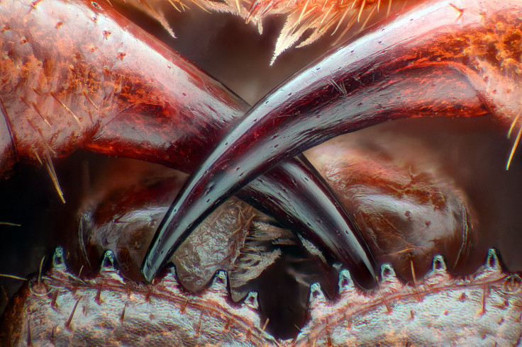
This image of the fearsome poison fangs of a centipede, magnified so that we can see every hair and battle scar, is one of the finalists in the annual Nikon Small World competition. Now in its 42<sup>nd year, the contest recognises excellence in photography taken under a microscope.
Walter Piorkowski, South Beloit, Illinois, USA: Poison fangs of a centipede (Lithobius erythrocephalus) – Fibre Optic Illumination/Image Stacking, 16xWalter Piorkowski
More than 2,000 photomicrography images from all over the world were entered into this year's competition. The judges have evaluated them on originality, informational content, technical proficiency and visual impact, and have narrowed the field down to 95 finalists.
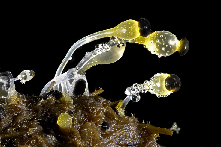
Many of the images reveal the hidden beauty that can be found in the most unlikely of places – such as this picture of delicate fungi growing on cow dung.
Michael Crutchley, Haverfordwest, Pembrokeshire, United Kingdom: Fungi on cow dung – Darkfield, 30xMichael Crutchley
This year, the public will select the winner of the Popular Vote category. You have until 25 October to choose your favourite image.
We've published 20 of our favourites in this gallery. You can see the other 75 finalists and vote for your winner at the Nikon Small World site.
Yousef Al Habshi, Abu Dhabi, United Arab Emirates: Eyes of a jumping spider (Hasarius adansoni) – Reflected Light, 9xYousef Al HabshiDr Igor Siwanowicz, Howard Hughes Medical Institute (HHMI), Janelia Research Campus, Ashburn, Virginia, USA: Front foot (tarsus) of a male diving beetle – Confocal, 100xDr Igor SiwanowiczJochen Schroeder, Chiang Mai, Thailand: Butterfly proboscis – Image Stacking, 6.3xJochen SchroederDr David Maitland, Feltwell, United Kingdom: Analgesic compound (salicin) extracted from Willow tree bark – Polarised Light, 50xDr David MaitlandDr Oscar Ruiz, the University of Texas MD Anderson Cancer Centre, Houston, Texas, USA: Four-day-old zebrafish embryo – Confocal, 10xDr Oscar RuizMatt Inman, Cremorne, New South Wales, Australia: Beta-alanine and taurine crystals – Transmitted Polarized Light/Focus Stacking, 10xMatt InmanDr Alvaro Roura, Instituto de Investigacións Mariñas,Ecology and Marine Biodiversity, Vigo, Spain: Viperfish (Chauliodus sloani) – Reflected Light/Darkfield, 20xDr Alvaro RouraMarek Mis, Suwalki, Podlaskie, Poland: Leg of a water boatman (Corixidae) – Polarized Light, Darkfield, 25xMarek MisDr Csaba Pintér, Keszthely, Zala, Hungary: Goatsbeard flower (Tragopogon orientalis) seeds – Focus Stacking, 1.4xDr Csaba PintérDouglas L Moore, University of Wisconsin - Stevens Point Museum of Natural History, Stevens Point, Wisconsin, USA: Polished slab of Teepee Canyon agate – Stereomicroscopy, 90xDouglas L MooreDavid Millard, Austin, Texas, USA: Curvepod Fumewort (Corydalis curvisiliqua) seed – Image Stacking, 4.5xDavid MillardRogelio Moreno Gill, Panama, Panama: Frontonia (single-celled organism) showing ingested food, cilia, mouth and trichocysts – Differential Interference Contrast, 200xRogelio Moreno GillLaurie Knight, Tonbridge, Kent, United Kingdom: Black elder tree flower stamen (Sambucus nigra) – Focus Stacking, 10xLaurie KnightKarl Gaff, Dublin Institute of Technology, School of Physics, Dublin, Ireland: CaptionMullein flower (Verbascum nigrum) – Reflected Light, 5xKarl GaffGeir Drange, Asker, Norway: Head section of an orange ladybird (Halyzia sedecimguttata) – Reflected Light/Focus Stacking, 10xGeir DrangeStefano Barone, Diatom Shop, Palazzo Pignano, Italy: 65 fossil Radiolarians (zooplankton) carefully arranged by hand in Victorian style – Darkfield, 100xStefano BaroneJose Almodovar, University of Puerto Rico, Mayaguez Campus, Biology Department, Mayaguez, Puerto Rico: Slime mould (Mixomicete) – Image Stacking/Reflected Light, 5xJose AlmodovarGeir Drange, Asker, Norway: Ant pupae (Myrmica sp.) – Reflected Light/Focus Stacking, 5xGeir Drange