Want to See What's Inside an Egyptian Mummy's Bandages? Now You Can Digitally Unwrap it

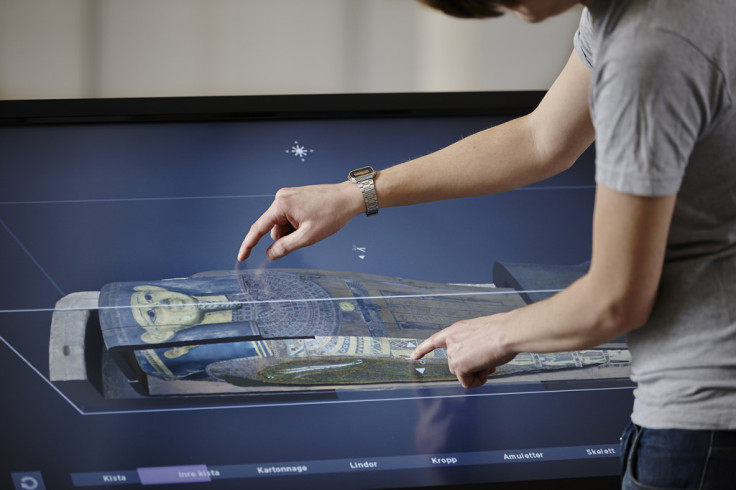
A Swedish museum is working on a virtual autopsy table that allows its visitors to unwrap Egyptian mummies and examine what's underneath.
Medelhavsmuseet, the Museum of Mediterranean and Near Eastern Antiquities in Stockholm owns eight Egyptian mummies, which have been digitally scanned using computed topography (CT) scanning.
The scans create density maps of the insides of the maps, which researchers from the Interactive Institute Swedish ICT have applied to a software platform they have developed called Inside Explorer.
Virtual autopsy table
Using the data from the CT scan and 2D photos of the coffins and mummy taken from multiple angles, Inside Explorer is able to build up an accurate 3D surface model using Autodesk Recap Photo software.

The finished product is a virtual autopsy table in an "embalmment room" next to the coffins, funerary goods and mummified remains of a wealthy Egyptian priest Neswaiu, who lived in the third century BC at the temple of the god Montu in Luxor.
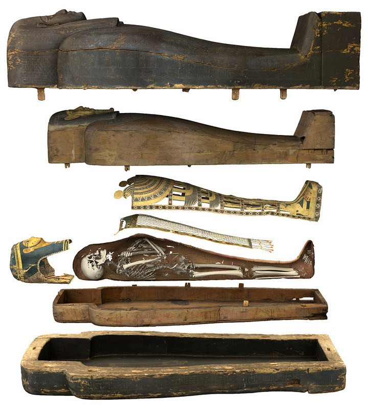
Museum visitors can use the touch screen to open Neswaiu's two coffins and then virtually pull back each layer of the mummy from the outermost layer down to his skeleton, and also view cross-sections of what the coffins and layers of mummy wrappings would have looked like.
When preparing him for burial, the embalmers took out Neswaiu's internal organs, wrapped them up, reinserted them and resealed the cut, placing amulets across each cut. Visitors can view the exact position of all 120 amulets that have been placed on the priest's body.
While the virtual autopsy table is already in use in numerous museums and science centres around the world, the software program for Neswaiu's mummy is the most complex autopsy table so far created.
"CT scanning gives you information about the interior of the mummy but it doesn't give you any colour or surface information," the research institute's Thomas Rydell told the BBC.
"So we continued the process by doing laser scanning and photogrammetry and that process gave us information about the surface and textures and colours of the mummy and then we're taking all that data and putting it on the table and making it accessible for museum visitors."
3D-printed amulet replica
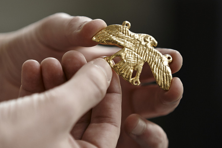
The CT scan is so powerful that a falcon-shaped amulet seen on Neswaiu's chest within the wrappings can be replicated without even moving it from the mummy.
The data from the scan is detailed enough that the researchers were able to 3D print a mould of the exact dimensions and details, and then cast an exact replica of the amulet.

The software platform was originally meant to help educate medical students and for use in hospitals, but the virtual autopsy table has proved so popular that many museums and science centres around the world have wanted to work with Interactive Institute Swedish ICT on interactive exhibits.
A virtual autopsy table was created for the British Museum's 5,500 year-old naturally mummified Egyptian Gebelein Man exhibit and was on display in 2013.
The system is also in use at museums and science centres in the USA, Sweden, Singapore, Spain, UK and Kazakhstan including the Smithsonian in Washington, London's Natural History Museum and the Field Museum of Natural History in Chicago.
© Copyright IBTimes 2025. All rights reserved.


